Why Spheres
Although your ultrasound machine QA protocol may indicate a proper calibration, the
existing presets can result in false negatives or positives, in both instances putting the
patient at risk or necessitating additional unnecessary testing.
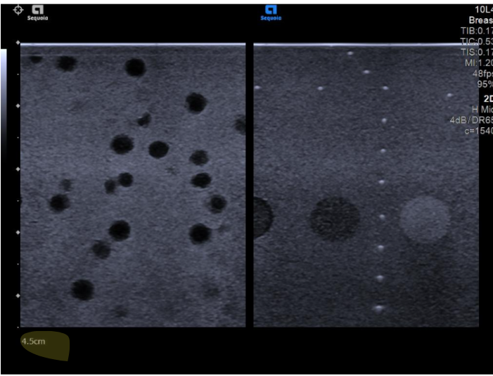
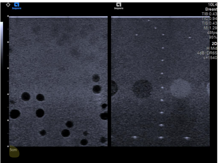
The difference between the two sets of images below is the depth of the Field of View
(FOV).
The first image has an FOV of 4.5 cm (default on the OEM Breast Preset), while the
second image has an FOV of 5.0 cm.
While the strings remain visible and unchanged between the two FOVs, the spheres in
the top half of the picture become verry blurry when the FOV is changed by as little as 5
mm.
The current QA protocols calibrate machines to the detection of strings. This means
that the image below would have passed the calibration test, despite completely missing
the spheres. By omitting a key element such as the detection of spheres, the current
QA protocols can lead to false diagnostics.
Risks of undetected dead zones
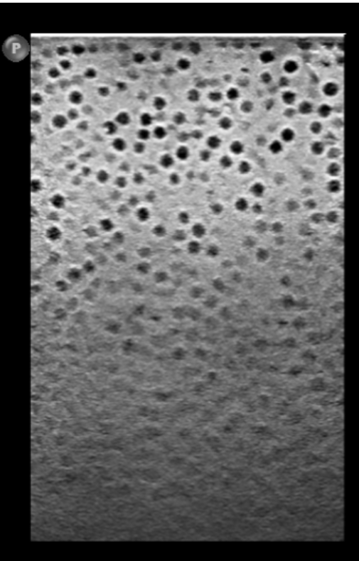
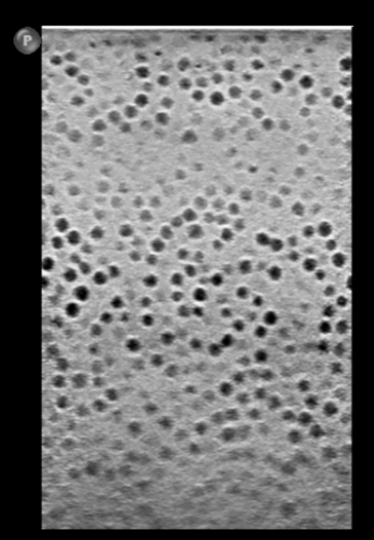
Can you notice the differences between the two images?
They are from the same machine, transducer, and obtained by the same technician.
One image has a Focal Zone of 2.5 - 7 cm, while the other has a Focal Zone of 0 - 5
cm.
Based on the Focal Zone presets, can you identify the area where there is a dead zone
that could result in an incorrect diagnosis?
Clinical Case
Here is a clinical scenario in which a physician is analyzing two images from the same
patient, each obtained with a different machine.
Both machines have the same OEM model, transducer, software presets, and were
operated by the same technician, scanning the same patient.
This first image showed indeterminate findings. This could have been labeled as a
negative or indeterminate test. Ultimately, the patient was asked to come back for a
repeat scan.
During the second scan, done with a different machine, the image had concerning
findings and the recommendation was made to proceed with a biopsy.
Fortunately, the radiologist caught the stark discrepancy between the two studies, and
when the OEM service team changed out the boards of the machines, it became
apparent that the calibration of the second machine had led to a false positive result,
which would have incorrectly put the patient through an unnecessary and invasive
procedure.
It is unclear how many patients have been scanned with the poorly calibrated machine.
However, we do know that Utune's Optimizer would have caught the issue with the
second machine as part of its calibration protocol.